
Effect of Yoga on Brain Functions
Effect of Yoga on Brain Functions
Scientific Perspective

Dear Readers,
Hope you had a good time learning about the effects of Yoga on structural/anatomical changes in brain (Give it a read if you haven’t). In this article (2/n), let’s delve further and learn more about the effects of Yoga on the functional and metabolic changes in the Brain.
The TL;DR*
Brain exhibits Neuroplasticity i.e. ability to change even as we grow older.
Studies have shown that yoga leads to functional changes in neural networks & brain metabolism which then drives
emotional regulation & reduced stress,
improved endurance to pain and
improved cognition (e.g. improved attention span & better memory).
While studies have been many the sample size is small and they also need to be updated for the post pandemic world of remote Yoga.
These studies have been done on yoga practioners and the results cited require long term committment and effort.

For those who love the details….
For decades it was believed that brain development is complete by the age of five and thus all the network of neural pathways were set and immutable. Now with digital imaging technology these myths have been shattered and now we understand and appreciate the concept of Neuroplasticity.
Technology and science work together, they are the gateways to invisible realms around us. Technology uses science to solve problems, and science uses technology to make new discoveries. Like with the invention of microscope and telescope, now we can see tiny microbes and faraway stars which earlier were not possible to see with the naked eye. In a similar fashion, with the advancement in neuroimaging techniques (functional MRI, diffusion tensor imaging, positron emission tomography) now we can examine and appreciate the innumerable effects of Yoga on neuroplasticity of Brain.
What we call reality is constantly shifting, as we expand our reach into the unknown - Anonymous
Effects of Yoga on Neural Activation and Functional Connectivity
In 1991, first ever PET scans1 were done on Yoga practitioners in meditative state and normal control state. Though the sample size was small (n=8), researchers found that the ratios of frontal to occipital metabolic rate of glucose was significantly elevated. Glucose metabolism in brain correlates with the activity in that region. To sum up the results, the functional brain scans of Yoga practitioners in meditative state demonstrated pronounced reduction in primary and secondary visual centers and slight increase of frontal region, indicating a holistic behaviour of the brain metabolism. Study conducted in 1998, demonstrated that during meditation brain activation pattern appears to be similar to brain during REM sleep and dreaming.
Later in 1999, another study2 examined the global cerebral blood flow (CBF) distribution in nine experienced subjects, differential regional CBF activity upon meditation was found in regions thought to support an executive attentional network. This study also reported the neural networks associated with the resting state of normal consciousness and the meditative state (See figure 1 and its legend for details). Later, Cohen and his team in 20093 further confirmed these findings in participants (n = 4) that underwent a 12-week Iyengar yoga training program. In coherence with these studies, several other research groups using perfusion fMRI demonstrated similar effects in the brains of practitioners during Kundalini yoga chanting meditation 4. Scientists observed strong positive correlations between depth of meditation and neural activity in the left inferior forebrain areas including the insula, inferior frontal cortex, and temporal pole. There were persistent changes in the left anterior insula and the precentral gyrus even after meditation was stopped5.
In simple words, these functional changes in neural networks help improve emotional regulation to reduce stress, anxiety and depression and additionally improves brain functioning.
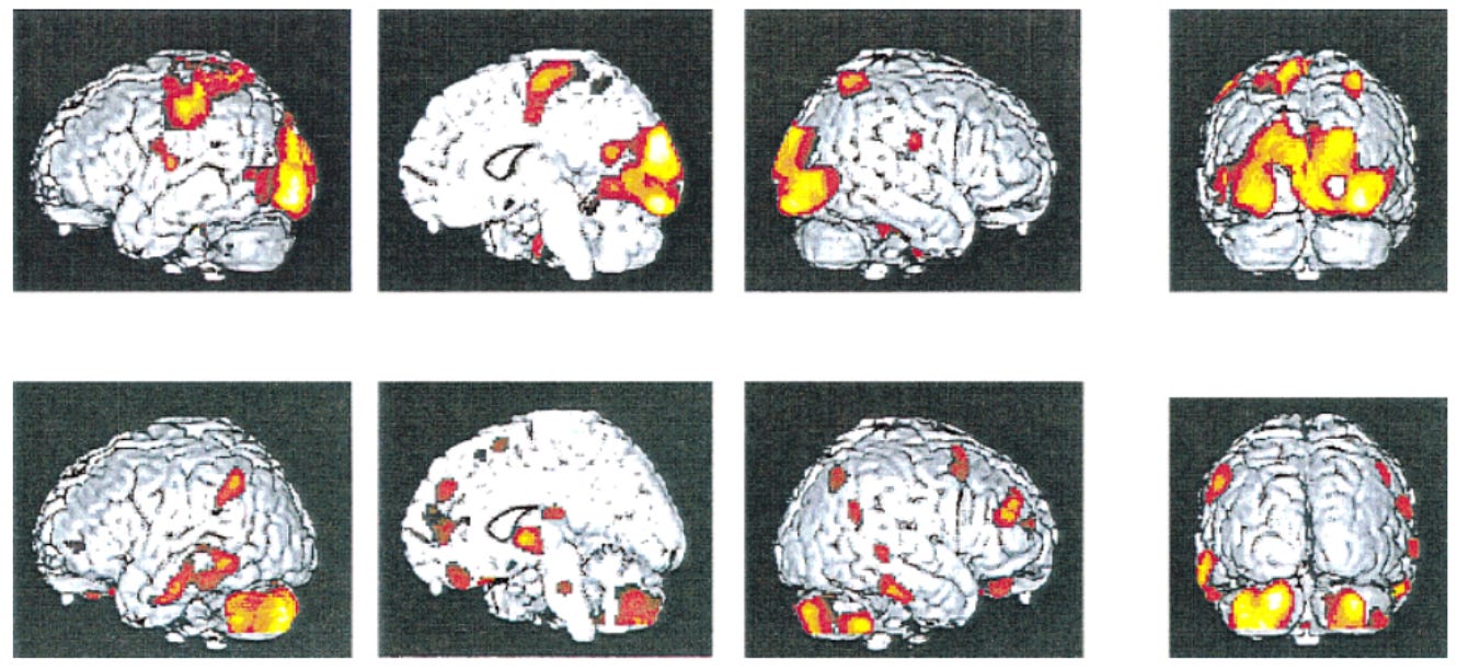
Figure 1: Cerebral activity patterns of combined meditative stages vs. conscious control (upper row) and vice-versa (lower row). The meditative state has differential activity mainly in anterior parietal and occipital regions. The conscious control demonstrated higher activity bilaterally in dorso-lateral orbital and cingulate frontal regions, posterior parietal region, temporal region, and the caudate nucleus, thalamus, pons, and cerebellar vermis and hemispheres [Adopted from Cohen et al, 2009].
Furthermore, multiple studies 6 7 8 9 showed differences in the activation or connectivity of the insula in yoga practitioners. The increased functional connectivity in the insular and frontal cortex of experienced yoga meditation practitioners simply relates to pain tolerance, better performance across a wide range of cognitive, emotional and motor skills. In addition, these studies further demonstrated significant deactivation of the right amygdala. The right hemisphere of the amygdala is associated with negative emotion. It plays a role in the expression of fear and in the processing of fear-inducing stimuli.
In 201810, one interesting study investigated the effects on emotional regulation in experienced yoga subjects (n = 19) compared to recreational athletes (n = 12) using emotionally arousing visual stimuli. When compared to athletes, brain scans of Yoga subjects depicted higher activation in the superior parietal lobule, postcentral gyrus, and anterior supramarginal gyrus during the emotion-evoking phase. These areas have been associated with attentional awareness and reduced egocentric bias.
Accordingly, individuals who practice yoga: (1) regulate the emotion generation process through greater flexibility, acceptance and non-attachment to the self when observing or experiencing emotions; and (2) be more empathetic when presented with an emotional situation, by attending to the emotions of other individuals.
Another study11 showed less activation in the dorsolateral pre-frontal cortex in yoga practitioners while performing a Sternberg working memory task. This result may point toward increased efficiency by experienced yoga practitioners while performing the task, which is in line with behavioural studies suggesting the positive influence of yoga on working memory performance.
If you are curious to understand more about the anatomy of brain and functional areas, refer to the figure 2 below:

Interestingly in 2015, Gard and colleagues12 performed a study to disentangle differences between yoga and meditation. Using Network-Based Statistics (NBS), which detects clusters of connections that significantly differ between groups, significant difference components for the comparison yoga practitioners (n=16) > controls (n=15) were found, comprising three nodes, with the caudate nucleus as the central node, connected to the parahippocampal gyrus and inferior temporal gyrus. Furthermore, compared to controls, meditators and yoga practitioners revealed equally stronger connectivity to a large number of brain regions.
Effects of Yoga on Brain Metabolites and Neurotransmitters
First magnetic resonance spectroscopy (MRS) study13 in yoga practitioners, demonstrated an increase in GABA levels while no changes were observed in the control subjects. In a subsequent interventional study, the same group measured MRS GABA levels in the left thalamus in healthy subjects that were randomly assigned to a 60-min yoga (n = 19) or walking (n = 15) intervention, three times a week for 12 weeks. The yoga subjects reported a greater improvement in mood and anxiety, compared to the walking group. Although no significant changes in thalamic GABA levels between groups were found, a significant positive correlation between changes in mood scales (revitalisation, tranquility, state-trait anxiety trait) and changes in thalamic GABA levels was found in the yoga group.
Another MRS study done on diabetic individuals14, that were assigned to either a yoga group (n = 34) or a control group (n = 34). The yoga group did yoga (including physical postures and breathing exercises) for six months, 6 days a week, for 45–60 min under daily supervision of a qualified yoga teacher. Results showed higher N-acetyl aspartate (is a potential promising biomarker of central neuronal dysfunction or loss) and lower myoinositol levels were found in the yoga group compared to the control group in the right dorsolateral frontal lobe, pointing toward higher neuronal integrity and lower neuroglial functioning, respectively.
In accumulation, these scientific studies are promising and consistently demonstrate structural changes in brain, increased neural connectivity followed with increased functioning and also positive metabolic changes in the brains of Yoga practitioners.
However, the number of studies is still limited and heterogeneous and several inconsistencies are present due to the heterogeneity among the different yoga styles included and the great variability in the applied research protocols. The integration of both neuroimaging and neurophysiological techniques (EEG, EMG, etc.) will further allow to investigate and bridge imaging findings with neurophysiological and behavioural assessment/improvements in well-being. Lastly, it remains to be determined whether web-based yoga interventions will be as effective as in-person yoga interventions which were primarily utilized in the reviewed papers.
Every man can, if he so desires, become the sculptor of his own brain -Santiago Ramon y Cajal (Spanish neuroscientist and pathologist)
Your mind and body are essentially rewiring when you practice yoga. Therefore, your attitudes, judgments, and inner dialogue are just as important as your breath and alignment when you practice.
I hope you enjoyed reading this article and after knowing how yoga can change your brain for good, it motivates you for daily practice! In the next article, I will cover the science behind the effect of Yoga on Endocrine System.
If you found the article of interest or have thoughts about what you’ve read here, please do share in the comments section below. It will help guide my future posts.
Herzog, H., Lele, V. R., Kuwert, T., Langen, K.-J., Kops, E. R., and Feinendegen, L. E. (1991). Changed pattern of regional glucose metabolism during yoga meditative relaxation. Neuropsychobiology 23, 182–187. [PubMed]
Lou, H. C., Kjaer, T. W., Friberg, L., Wildschiodtz, G., Holm, S., and Nowak, M. (1999). A15O-H2O PET study of meditation and the resting state of normal consciousness. Hum. Brain Mapp. 7, 98–105 [Google Scholar]
Cohen, D. L., Wintering, N., Tolles, V., Townsend, R. R., Farrar, J. T., Galantino, M. L., et al. (2009). Cerebral blood flow effects of yoga training: preliminary evaluation of 4 cases. J. Altern. Complement. Med. 15, 9–14. [PubMed]
Khalsa, S. B., Cohen, L., McCall, T., and Telles, S. (2016). The principles and practice of yoga in health care. Int. J. Yoga 11, 86–87. [Google Scholar]
Wang, D. J. J., Rao, H., Korczykowski, M., Wintering, N., Pluta, J., Singh, D., et al. (2011). Cerebral blood flow changes associated with different meditation practices and perceived depth of meditation. Psychiatry Res. Neuroimaging 191, 60–67. [PubMed]
Kalyani B. G., Venkatasubramanian G., Arasappa R., Rao N. P., Kalmady S. V., Behere R. V., et al. (2011). Neurohemodynamic correlates of “OM” chanting: a pilot functional magnetic resonance imaging study. Int. J. Yoga 4 3–6. [PubMed]
Froeliger B., Garland E. L., McClernon F. J. (2012). Yoga meditation practitioners exhibit greater gray matter volume and fewer reported cognitive failures: results of a preliminary voxel-based morphometric analysis. Evid. Based Complement. Alternat. Med. 2012 1–8. [PubMed]
Hernández S. E., Barros-Loscertales A., Xiao Y., González-Mora J. L., Rubia K. (2018). Gray matter and functional connectivity in anterior cingulate cortex are associated with the state of mental silence during Sahaja yoga meditation. Neuroscience 371 395–406. [PubMed]
Hernández S. E., Suero J., Barros A., González-Mora J. L., Rubia K. (2016). Increased grey matter associated with long-term Sahaja yoga meditation: a voxel-based morphometry study. PLoS One11:e0150757. [PubMed]
Wadden, K. P., Snow, N. J., Sande, P., Slawson, S., Waller, T., and Boyd, L. A. (2018). Yoga practitioners uniquely activate the superior parietal lobule and supramarginal gyrus during emotion regulation. Front. Integr. Neurosci. 12:60. [PubMed]
Gothe, N. P., Hayes, J. M., Temali, C., and Damoiseaux, J. S. (2018). Differences in brain structure and function among yoga practitioners and controls. Front. Integr. Neurosci.12:26. [PubMed]
Gard, T., Taquet, M., Dixit, R., Hölzel, B. K., Dickerson, B. C., and Lazar, S. W. (2015). Greater widespread functional connectivity of the caudate in older adults who practice kripalu yoga and vipassana meditation than in controls. Front. Hum. Neurosci. 9:137. [PubMed]